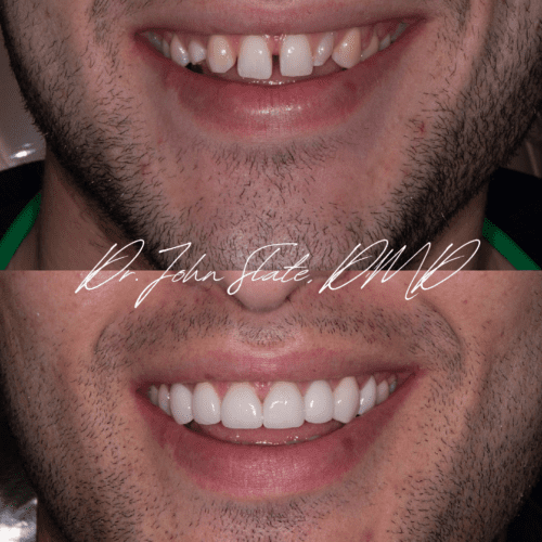
This patient presented with a congenitally missing lateral tooth #7 and a congenital peg lateral on tooth #10. He also had retained his adolescent/primary tooth “C” taking the place of his canine position, which had moved into the congenitally missing #7 position. This treatment was a multidisciplinary treatment plan involving our local orthodontist and periodontist. He wanted to correct his smile, something he had always been self conscious about, and couldn’t be happier with his final result!
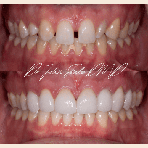
This patient presented with a congenitally missing lateral tooth #7 and a congenital peg lateral on tooth #10. He also had retained his adolescent/primary tooth “C” taking the place of his canine position, which had moved into the congenitally missing #7 position. This treatment was a multidisciplinary treatment plan involving our local orthodontist and periodontist. He wanted to correct his smile, something he had always been self conscious about, and couldn’t be happier with his final result!
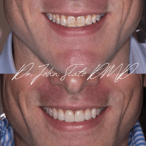
This patient was looking for cosmetic treatment to treat the developmental white spot lesions which were visibly apparent on his front teeth. We aimed to recreate the exact shape and anatomy of his teeth, but with porcelain veneers to mask the discoloration of his natural teeth just below the surface. This transformation took about 2-3 appointments to deliver.
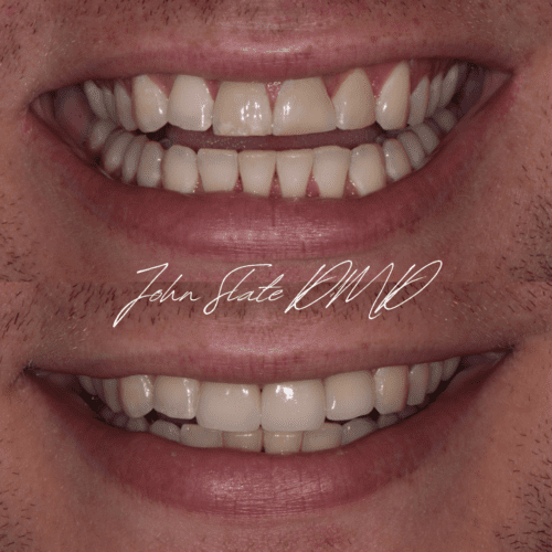
This patient was in an accident when he was younger, causing a fracture and nerve death on 3 of his upper front teeth. You can notice flattening and wear on the edges of his anterior teeth, which continue to chip and break down over time. We eventually decided to treat these teeth with a more predictable treatment, to avoid continued breakdown and cosmetic issues. Teeth #8, 9, and 10 were previously root canal treated, and restored with full coverage porcelain crowns.
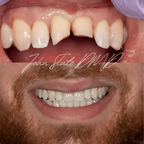
This patient tripped and fell, fracturing his anterior 2 teeth in half. His initial treatment began in an emergency room, and the remainder of his care was completed in our office. The unaesthetic “emergency” composite bonding was removed and both teeth #8 and #9 were restored with a core-build up and porcelain veneers. We were able to keep the cingulum intact on the tongue side of the tooth to maintain the overall strength of these central incisors. We will monitor these teeth in the long term for any nerve damage caused by his accident.
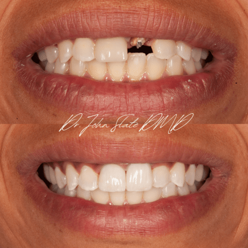
This patient had an accident when younger, causing damage to her anterior 2 central incisors. They were historically treated with root canal therapy and a post was placed to help restore the teeth. Both root canals treatments remained successful, but the post on tooth #9 was an inadequate length for retention of the build-up and crown. She presented to our office with the crown and build-up in hand. We planned for removal of the post on #9 and replacement with a longer, more retentive post and core build-up. Prior to finalizing the shade selection for these crowns, this patient whitened her teeth, and we matched the porcelain shade to her new tooth color. For symmetry we replaced the crown on tooth #8 to deliver this healthy and beautifully restored smile!
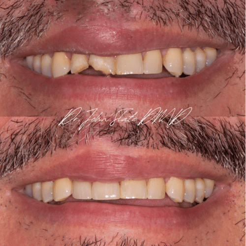
This patient was in a biking accident in DC causing damage to his lateral and central incisor. The teeth were splinted to his adjacent teeth, allowing the bone to heal following his accident. The nerves were severed on these teeth during the accident, so the teeth were treated with root canal therapy before restoring with core build-ups and full coverage porcelain crowns. It is often very challenging to match the symmetrical teeth shades, and we were extremely pleased with his final cosmetic result.
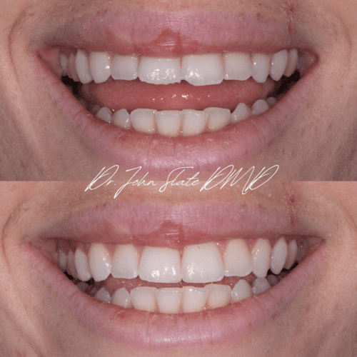
This patient had some minor chipping of her central incisors after getting bumped in the elbow while drinking from a bottle. This treatment took under an hour to restore the incisal edges of #8 and #9 with composite bonding in a single appointment.
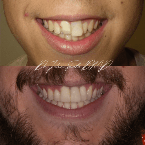
This patient’s treatment was caused by a baseball accident at the age of 9. The tooth was completely avulsed from the mouth, and reimplanted by his pediatric dentist. The tooth was root canal treated at the time, and was not monitored by any dentist afterwards for close to a decade. During that time, the tooth underwent a process called external resorption, where the body began to reject the re-implanted tooth causing significant damage to the surrounding bone and gum tissues. He presented to our office for multi-disciplinary treatment involving extraction of tooth #9, a very large bone graft, and implant therapy. Ultimately we restored teeth #7, 8 and 10 with full coverage crowns, to allow space for an implant and implant crown on tooth #9.
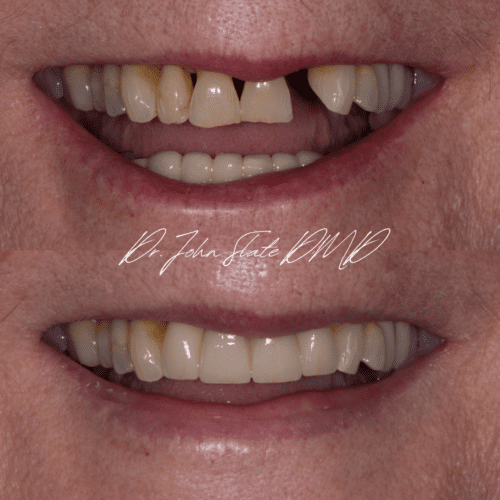
This elderly patient presented with a fractured tooth #10 which was non-restorable. The patient requested treatment to replace tooth #10, but also complained about the “black triangles” near the gum line between her teeth. There was minimal remaining bone surrounding teeth #7-10 and mobility of the teeth due to past periodontal disease. We planned to remove teeth #7-10, restore with two implants in the #7 and #10 tooth position, and deliver a 4-unit Zirconia (ceramic) bridge to restore teeth #7, 8, 9 and 10.
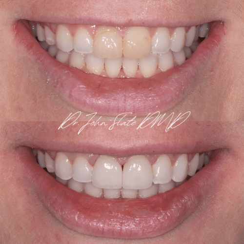
This patient had an accident when younger, damaging both of her central incisors. In dentistry, we usually will not restore teeth with veneers or crowns until the patient's jaw bone has finished growing. As the jaw bones grow, the teeth can shift and the gum line position can change, which can be problematic for the transition line between the porcelain and tooth. She had just finished orthodontic treatment, was finished growing, and was ready for a more definitive cosmetic result. We restored both central incisors with porcelain veneers on #8 and #9.

This patient presented to our office seeking a cosmetic treatment to replace his older composite veneers on his central incisors. Both teeth were healthy, and we treated #8 and #9 with porcelain veneers to completely restore the form, color, and function of his teeth.
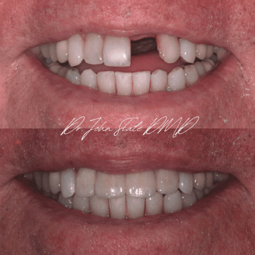
This patient presented after his central incisor had fractured off to the gum line. Tooth #9 was non-restorable and needed to be removed. The patient had to make a decision to restore tooth #9 with a single implant or a 4-unit bridge. He chose to restore teeth #7-10 with a PFM bridge to strengthen his adjacent teeth, and provide support for his floating crown in the #9 position. He was able to restore much quicker with this treatment option and was also able to avoid additional grafting and implant surgery which would be required for successful implant therapy.
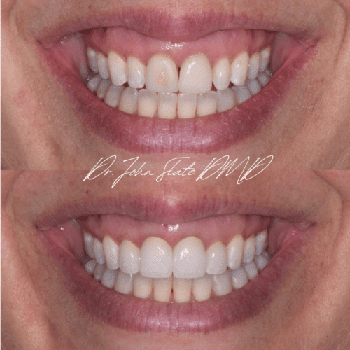
This patient had crowns placed on teeth #8 and #9 by another local dentist. When the crowns were benign delivered, the porcelain was overly thinned on the facial aspect of these crowns. After the adjustment the core or dentin of the tooth was showing right through the center of the crown. The patient was seeking a more esthetically pleasing result, and also was concerned with the appearance of her narrow lateral incisors. We planned to restore teeth #7 and #10 with porcelain veneers and the crowns on teeth #8 and #9 were replaced to match the color, form, and function of her maxillary incisors.

This treatment showcases a common treatment for abfractions of class V cervical lesions. The neckline of teeth #5, 6, 7, 10, and 11 had worn over time due to clenching and grinding, creating small divots on the facial aspect of the teeth. These areas were an esthetic issue for the patient, and oftentimes would become food traps when eating. This treatment involved a single appointment to restore the facial aspect of these teeth with composite bonding in under an hour and a half.
